ICU Mobilisation: Evidence, Safety Screening and Progression Criteria
Learn ICU mobilisation using Hodgson red-yellow-green safety criteria, ABCDEF bundle context, progression from passive ROM to ambulation, and ICU-acquired weakness prevention.
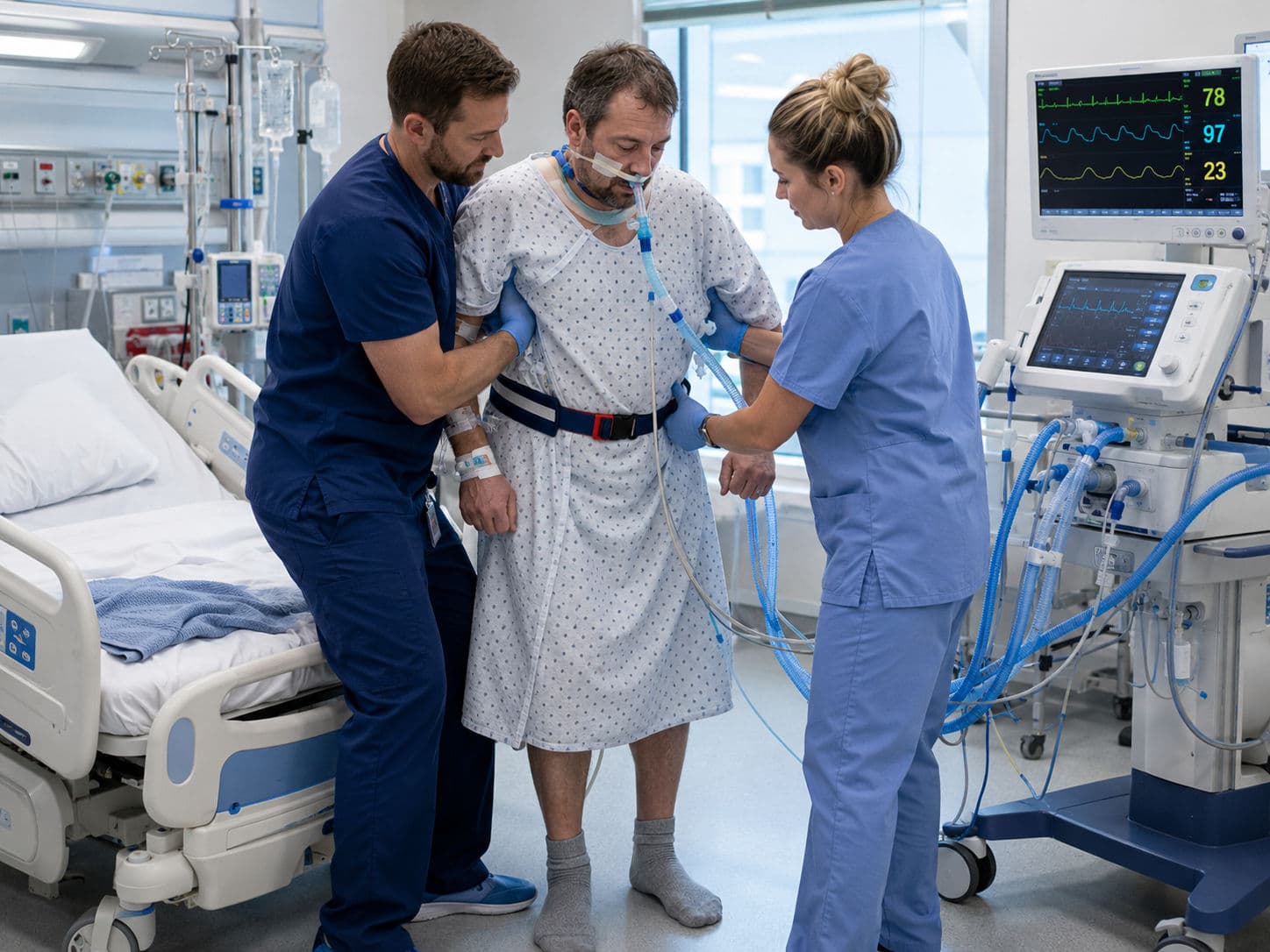
ICU mobilisation is no longer viewed as an optional “extra” after the patient becomes fully stable. In modern critical care, movement is part of treatment. For physiotherapists, the clinical challenge is to start early enough to prevent deconditioning but safely enough to avoid respiratory, cardiovascular or line-related complications. ICU mobilisation may include passive range of motion, active exercises in bed, sitting over the edge of the bed, standing, transferring to a chair and ambulation with appropriate monitoring.
The aim is not simply to make the patient walk. The aim is to preserve muscle function, improve ventilation, reduce immobility-related complications, support delirium prevention and prepare the patient for functional recovery after ICU discharge. This is especially important because prolonged ventilation, sepsis, inflammation, corticosteroid exposure, neuromuscular blockade, hyperglycaemia and bed rest contribute to intensive care unit-acquired weakness, or ICU-AW (Fan et al., 2014; Jolley et al., 2016).
Why ICU Mobilisation Matters
Critical illness rapidly affects skeletal muscle. Muscle wasting can begin within the first week of ICU admission, especially in mechanically ventilated or septic patients. The diaphragm is also affected because mechanical ventilation unloads respiratory muscles. The result may be limb weakness, poor cough, reduced exercise tolerance, difficulty weaning from ventilation and delayed independence in activities of daily living.
Early physiotherapy interrupts this cycle. Passive and active movement maintains joint mobility, stimulates circulation and provides sensory input. Sitting and standing increase antigravity muscle activity, improve ventilation-perfusion matching and help the patient relearn upright tolerance. Ambulation, when safe, restores task-specific function. Classic cardiopulmonary physiotherapy texts, including Cash, Hillegass, and Frownfelter and Dean, emphasise that ICU rehabilitation must be based on oxygen transport, haemodynamic response, ventilatory reserve and functional need rather than bed exercises alone (Frownfelter et al., 2022; Hillegass, 2022; Webber & Pryor, 1993).
The evidence is supportive but nuanced. Earlier trials showed that early physical and occupational therapy during mechanical ventilation improved functional independence and reduced delirium duration (Schweickert et al., 2009). Systematic reviews suggest that early rehabilitation can reduce the likelihood of ICU-AW (Anekwe et al., 2020). However, the large TEAM trial found that a higher-dose early active mobilisation strategy during mechanical ventilation did not improve days alive and out of hospital at 180 days compared with usual care and was associated with more adverse events (Hodgson et al., 2022). Therefore, the exam-ready conclusion is: ICU mobilisation is beneficial when it is early, screened, progressive and individualised—not when intensity is pushed blindly.
ICU Mobilisation in the ABCDEF Bundle
The ABCDEF bundle gives ICU mobilisation its broader clinical context. It includes:
A — Assess, prevent and manage pain
B — Both spontaneous awakening trials and spontaneous breathing trials
C — Choice of analgesia and sedation
D — Delirium assessment, prevention and management
E — Early mobility and exercise
F — Family engagement and empowerment
For physiotherapists, the “E” element cannot work in isolation. A deeply sedated, painful or delirious patient may not participate safely. Similarly, a patient who has not had appropriate ventilator, haemodynamic or line review may be unsafe to mobilise. Therefore, ICU mobilisation depends on multidisciplinary coordination with the intensivist, nurse, respiratory therapist and, where appropriate, the family.
In practice, the physiotherapist should ask the following: Is pain controlled? Is sedation light enough? Is oxygenation stable? Are vasoactive drugs stable or escalating? Is the airway secure? Are invasive lines protected? Is the patient able to follow commands, or can the team safely assist despite delirium? This approach converts mobilisation from a risky bedside event into a structured ICU intervention.
Safety Screening: Hodgson Red, Yellow and Green Criteria
Hodgson and colleagues proposed a traffic-light approach for active mobilisation of mechanically ventilated ICU patients. The framework considers respiratory, cardiovascular, neurological and other medical-surgical factors (Hodgson et al., 2014). It is not meant to replace local ICU protocols, but it gives physiotherapists a practical safety language.

Respiratory green signs usually include a secure airway, acceptable ventilator synchrony, SpO₂ generally ≥90%, FiO₂ ≤0.6, respiratory rate below about 30 breaths/min and no rapidly escalating oxygen or ventilatory support. Yellow signs include borderline oxygenation, higher respiratory rate, increased work of breathing or recent changes in ventilator settings. Red signs include severe desaturation despite support, marked respiratory distress, an unsafe airway, uncontrolled ventilator asynchrony or a need for rescue therapies such as prone ventilation or unstable high ventilatory support.
Cardiovascular green signs include a stable rhythm, acceptable blood pressure, no active myocardial ischaemia and stable or minimal vasoactive support. Yellow signs include controlled arrhythmias, borderline blood pressure or stable low-dose vasopressors where the team agrees mobilisation is reasonable. Red signs include new unstable arrhythmia, active chest pain or ischaemia, uncontrolled hypotension or hypertension, rising lactate with shock, or escalating vasoactive support.
Neurological screening includes consciousness, cooperation and safety. A calm patient who follows commands is usually safer for active mobilisation. Delirium does not automatically prohibit mobilisation, but agitation, inability to protect lines or reduced consciousness may require modification or deferral. Other red flags include active bleeding, unstable fractures, open surgical wounds requiring restriction, uncontrolled intracranial pressure, unstable spinal precautions, or femoral lines/sheaths where local policy limits hip movement.
Progression of ICU Mobility
ICU mobilisation should progress from low-load movement to functional, task-specific activity. The level chosen depends on screening, baseline function, current physiological reserve and goals of care.
The first level is positioning and passive range of motion. This is used for deeply sedated, unconscious or very weak patients. It helps prevent stiffness, pressure injury and dependent atelectasis, but it is not enough once the patient can actively participate.
The second level is active-assisted and active bed exercise. The patient performs ankle pumps, heel slides, upper-limb movements, bridging, rolling and breathing control. This stage is useful when the patient is awake but not yet ready for upright activity.
The third level is sitting. This may begin with head-up positioning, then chair positioning in bed, then sitting over the edge of the bed. Sitting challenges trunk control, improves lung expansion and gives an early measure of orthostatic tolerance.
The fourth level is a sit-out or transfer to a chair. This is a major functional milestone because it loads the lower limbs, stimulates alertness and improves tolerance to upright posture. Transfers may require a hoist, tilt table, standing aid, multiple staff members or ventilator-portable equipment.
The fifth level is standing and marching. The physiotherapist assesses lower-limb strength, balance, blood pressure response, respiratory effort and fatigue. Standing may be static initially, followed by weight shifts, mini-squats or marching on the spot.
The sixth level is ambulation. Walking may be done with a walker, portable monitor, portable ventilator or oxygen support and adequate staff for airway, lines and balance. The first walk may only be a few steps, but it marks a transition from impairment-based treatment to functional rehabilitation.

Monitoring, Stopping Rules and ICU-AW Prevention
During mobilisation, monitor SpO₂, respiratory rate, heart rate, blood pressure, rhythm, work of breathing, pain, fatigue, consciousness and patient appearance. In ventilated patients, also observe ventilator synchrony, airway security, tubing tension and alarm trends. Stop or downgrade the session if there is marked desaturation, severe breathlessness, new chest pain, dizziness, syncope, unstable arrhythmia, excessive blood pressure change, line dislodgement, patient distress or a sudden neurological change.
ICU-AW prevention requires more than exercise alone. The physiotherapy role includes early screening for weakness, progressive activity, positioning, breathing exercises, airway clearance when indicated, inspiratory muscle consideration during ventilator weaning, and education after ICU discharge. The wider ICU team contributes through sedation minimisation, glycaemic control, nutrition, sepsis management and avoiding unnecessary immobilisation. A simple bedside tool such as the Medical Research Council sum score can help identify clinically significant weakness when the patient is awake and cooperative; a score below 48/60 is commonly used to support ICU-AW diagnosis (Fan et al., 2014).
One-Glance ICU Mobilisation Progression Algorithm
Step 1: Daily readiness screen
Review respiratory, cardiovascular, neurological and medical-surgical safety.
Step 2: Assign traffic light
Green = proceed.
Yellow = modify and discuss.
Red = defer, treat instability and reassess.
Step 3: Choose highest safe level
Passive ROM → active bed exercise → sitting in bed → edge-of-bed sitting → sit-out → standing → marching → ambulation.
Step 4: Monitor continuously
SpO₂, respiratory rate, heart rate, blood pressure, rhythm, symptoms, airway, ventilator tubing and lines.
Step 5: Progress, pause or stop
Progress if stable.
Pause if borderline.
Stop if a red-flag response appears.
Step 6: Document functionally
Record mobility level, assistance required, physiological response, barriers and next-session goal.

Viva-Ready Summary
ICU mobilisation is an evidence-based, multidisciplinary intervention used to reduce immobility complications and support recovery in critically ill patients. It should be delivered within the ABCDEF bundle and guided by safety screening. Hodgson’s red-yellow-green approach helps physiotherapists decide whether to proceed, modify or defer mobilisation. Progression begins with positioning and passive range of motion, then advances to active exercise, sitting, sit-out, standing and ambulation. The current evidence supports early, individualised mobilisation, but not aggressive high-intensity mobilisation for every ventilated patient.
One-line recall point:
ICU mobilisation means “screen first, mobilise early, progress functionally and stop when physiology says stop".
References
Anekwe, D. E., Biswas, S., Bussières, A., & Spahija, J. (2020). Early rehabilitation reduces the likelihood of developing intensive care unit-acquired weakness: A systematic review and meta-analysis. Physiotherapy, 107, 1–10. doi: 10.1016/j.physio.2019.12.004
Fan, E., Cheek, F., Chlan, L., Gosselink, R., Hart, N., Herridge, M. S., Hopkins, R. O., Hough, C. L., Kress, J. P., Latronico, N., Moss, M., Needham, D. M., Rich, M. M., Stevens, R. D., Wilson, K. C., & Winkelman, C. (2014). An official American Thoracic Society clinical practice guideline: The diagnosis of intensive care unit-acquired weakness in adults. American Journal of Respiratory and Critical Care Medicine, 190(12), 1437–1446. doi: 10.1164/rccm.201411-2011ST
Frownfelter, D., Dean, E., Stout, M., Kruger, R., & Anthony, J. (2022). Cardiovascular and pulmonary physical therapy: Evidence and practice (6th ed.). Elsevier.
Hillegass, E. (2022). Essentials of cardiopulmonary physical therapy (5th ed.). Elsevier.
Hodgson, C. L., Stiller, K., Needham, D. M., Tipping, C. J., Harrold, M., Baldwin, C. E., et al. (2014). Expert consensus and recommendations on safety criteria for active mobilization of mechanically ventilated critically ill adults. Critical Care, 18, Article 658. doi: 10.1186/s13054-014-0658-y
Hodgson, C. L., Bailey, M., Bellomo, R., Brickell, K., Broadley, T., Buhr, H., et al. (2022). Early active mobilization during mechanical ventilation in the ICU. New England Journal of Medicine, 387(19), 1747–1758. doi: 10.1056/NEJMoa2209083
Jolley, S. E., Bunnell, A. E., & Hough, C. L. (2016). ICU-acquired weakness. Chest, 150(5), 1129–1140. doi: 10.1016/j.chest.2016.03.045
Schweickert, W. D., Pohlman, M. C., Pohlman, A. S., Nigos, C., Pawlik, A. J., Esbrook, C. L., et al. (2009). Early physical and occupational therapy in mechanically ventilated, critically ill patients: A randomised controlled trial. The Lancet, 373(9678), 1874–1882. doi: 10.1016/S0140-6736(09)60658-9
Webber, B. A., & Pryor, J. A. (1993). Cash’s textbook of chest, heart and vascular disorders for physiotherapists (4th ed.). Faber & Faber.